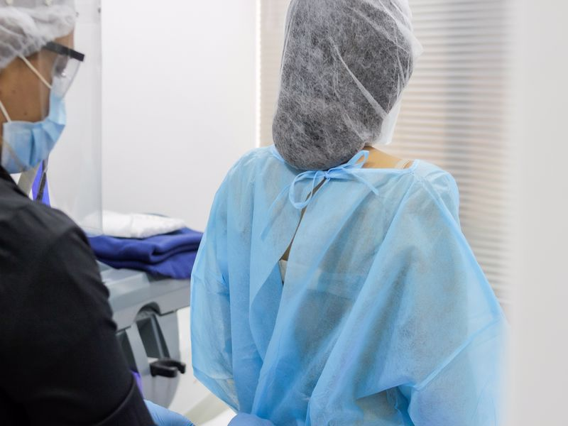
Breast cancer diagnosis includes some self-check methods that individuals can perform. Every woman over the age of 40 should regularly undergo a mammogram. This process should be repeated every two years. Individuals under the age of 40 can examine themselves through self-examination techniques.
Contents
Self-Examination for Breast Cancer Diagnosis
Self-examination for breast cancer diagnosis can be performed. Starting at the age of 20, individuals can perform this examination a few days after the end of their menstrual cycle. It should be done once a month. The self-examination method is as follows:
- Stand in front of a mirror.
- The environment should have sufficient lighting.
- Place hands on the waist and observe whether both breasts are the same.
- Check for differences in color, shape, or size between the two breasts.
- Then place hands behind the head.
- Observe again to see if there are any differences.


How to Perform Self-Examination for Breast Cancer Diagnosis?
Self-examination for breast cancer diagnosis also includes additional steps. After the check described above, the following steps can be performed a few days after the menstrual period ends:
- Lie on your back.
- Use your left hand to examine the right breast.
- Use your right hand to examine the left breast.
- Make small circular motions over each breast.
- Press lightly, moving from top to bottom and bottom to top.
- Check for any swelling or hardness.
- After examining the breasts, also check the underarm areas.
Doctor's Examination in Breast Cancer Diagnosis
The role of the doctor is crucial in breast cancer diagnosis. After self-examination, if a lump is detected, the individual should consult a doctor. The doctor carefully examines both breasts and underarms. Following the physical examination, the doctor directs the patient for tests and further evaluations.
Imaging Methods in Breast Cancer Diagnosis
Imaging methods used in breast cancer diagnosis are as follows:
- Mammography: This involves taking X-ray images of breast tissue. During the procedure, the breast is placed between two plates of a mammography device. A small amount of compression is applied, and a low dose of X-rays is used to capture images of the breast tissue. The images are transferred to a computer to determine the presence of any lumps.
- Ultrasound: One of the most frequently used methods in breast imaging. Ultrasound is especially useful for examining suspicious lesions detected in mammography and for performing biopsies when necessary. It is also used for evaluating regional lymph nodes.
- MRI: Used particularly for women with dense breast tissue and suspicious lesions identified in mammography during breast cancer investigations. It can also evaluate the other breast in women already diagnosed with cancer in one breast. MRI is a method used for screening high-risk women, such as those with genetic mutations or a history of chest radiation exposure.


Biopsy Methods in Breast Cancer Diagnosis
Biopsy methods used in breast cancer diagnosis are as follows:
- Fine-Needle Aspiration Biopsy: Evaluates lesions detected in the breast at the cellular level. It is used to distinguish between benign and malignant conditions. It is also one of the critical parameters in determining breast cancer stages and assessing regional lymph node metastasis.
- Excisional Biopsy: A more invasive method that can be used when core needle biopsy cannot be performed or when pathological results are inconsistent with imaging findings. It is also used for lesions that might be confused with cancer and is required in cases of suspected phyllodes tumors.
- Vacuum Biopsy: Used only for lesions visible on mammography. Since these lesions often carry a risk of breast cancer, they are removed using this method. Benign small lumps may also be removed using this method to eliminate potential risks.


